

The One Question to Ask Yourself About Every Histology Slide
Ask it before you try to identify anything — and watch your study time get cut in half
Most vet students approach histology the same way. They open a slide, squint at it, flip to their notes, look for something that matches, and hope the pattern sticks. Sometimes it does. Usually it does not. They flip back to the slide. They squint some more. They move on with a vague sense that they sort of know what that tissue is and a growing certainty that they definitely do not.
The problem is not the slide. The problem is the order of operations.
Before you try to identify a cell type, a tissue, or an organ, there is one question that should come first. Always. Every single slide. It is not a trick or a shortcut. It is the question that trained histologists ask automatically, the one experienced pathologists have internalized so deeply they do not even notice they are asking it anymore. Once you start asking it consciously, you will wonder how you ever looked at slides without it.
The question is this: “What is this tissue’s job?”
Ask it before you look for anything else. Before you identify cells. Before you match anything to your notes. Before you even adjust the magnification.
That is it. That is the whole article. Everything that follows is just explaining why it works so you will actually do it.
Why “What Does It Look Like?” Is the Wrong First Question
When you approach a slide by looking for visual features first — the color, the shape, the staining pattern — you are starting with the output and working backward to the meaning. That is the hard direction. It requires your brain to hold a large number of visual possibilities simultaneously and compare the slide against each one. This is cognitively expensive and produces unreliable results because one tissue can look startlingly similar to another when you do not know what you are looking for.
Histology is not a visual memorization subject. It is a functional anatomy subject that happens to use visual tools. Every structure on a slide exists because some cell or tissue has a job to do, and that job determines everything about how the tissue is built. The architecture is not arbitrary. It is the direct physical expression of function.
When you start with function, you constrain the problem. You are not comparing your slide to every tissue you have ever studied. You are asking a much smaller question: given what this tissue is supposed to do, what kind of structure would evolution have built to do it? Then you look at the slide to confirm or revise your hypothesis. That is the forward direction, and it is dramatically faster.
Histology is not a visual memorization subject. It is a functional anatomy subject that happens to use visual tools. Start with function and the structure follows logically.
The Framework: Four Jobs, Four Structures
Virtually every tissue you will encounter in first-year histology has one of four primary jobs. The job tells you what to expect before you look at a single cell.
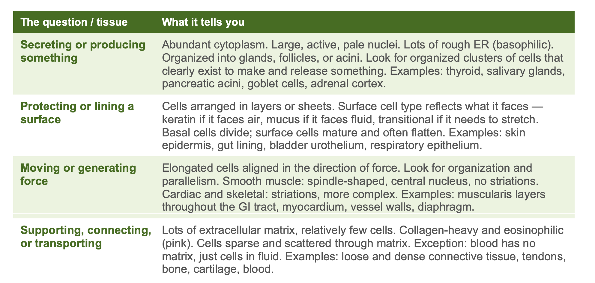
Those four categories cover the majority of what you will see in the first two years. When you open a slide, your first question is which category fits. Everything after that is narrowing within the category, not searching through all of histology at once.
A Walk-Through: Small Intestine
Here is what this looks like in practice, using small intestinal mucosa as the example because you will see it on every histology exam you take in year one.
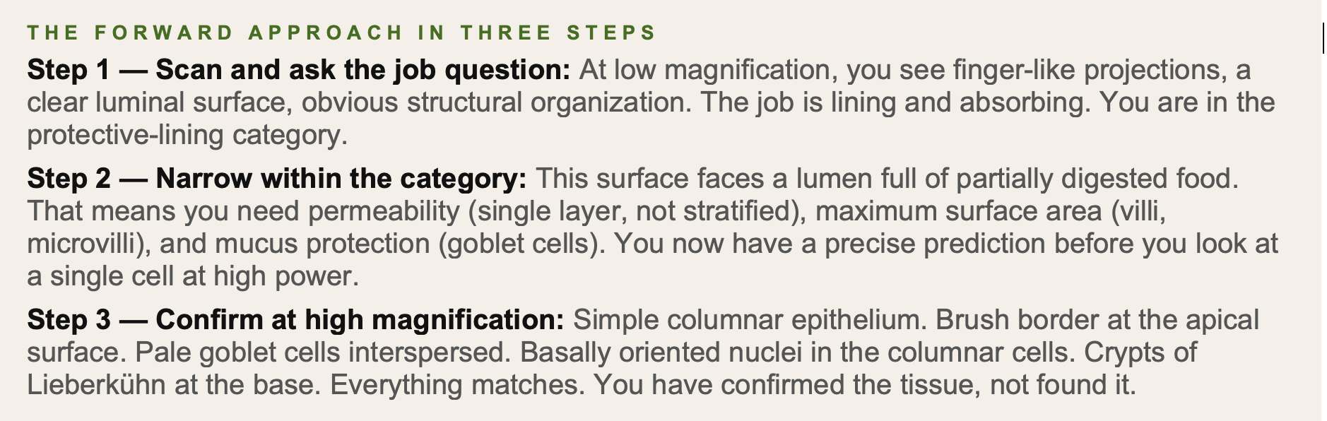
The difference between confirming and finding sounds subtle. It is not. Confirming takes about ten seconds. Finding — the unfocused searching approach — can take several minutes and often ends in a wrong answer because your eye gets drawn to irrelevant features.
Confirming takes ten seconds. Finding takes several minutes and often ends in the wrong answer. The job question converts every slide from a mystery into a confirmation exercise.
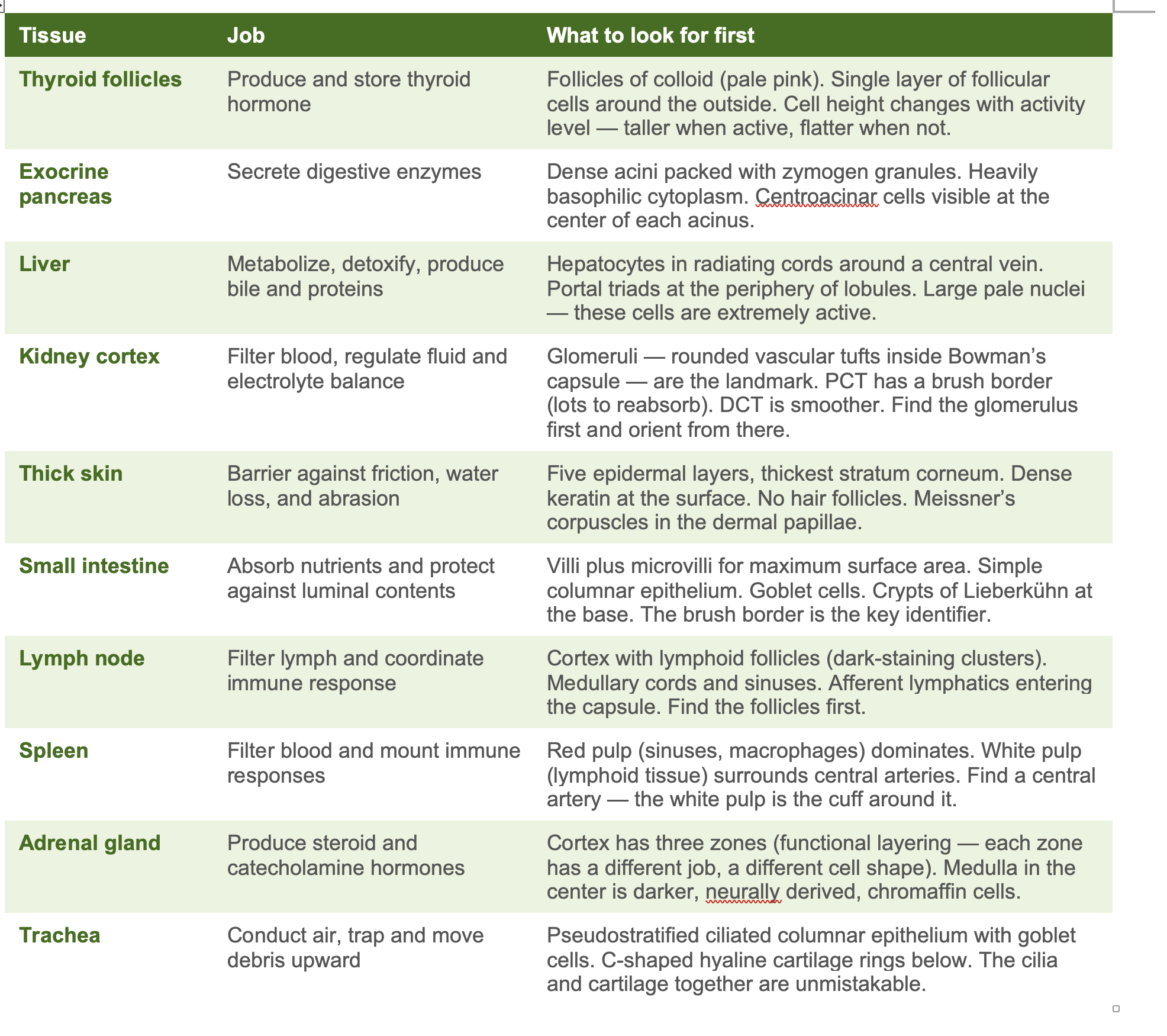
The Tissue Job Reference Table — Print This, Stick It Up
Here is the same logic applied across the tissues you will see most often in year one. Before lab and before exams, scan this table and ask the job question for each tissue before you look at any images.
The Magnification Sequence That Locks It In
The job question works best when you pair it with a consistent magnification sequence. Most students jump straight to high magnification, which is like trying to read a map by zooming into a single intersection. You lose the context that reveals functional organization.
SCAN → SURVEY → CONFIRM
Scan (lowest power): Ask the job question here. What is the overall architecture? Glands? Layers? Tubes? Follicles? Cords? This is where you make your functional category call. Do not skip this step.
Survey (medium power): Narrow within the category. What type of epithelium? What is the cell shape and arrangement? More pink or more purple, and where? Read the nuclei — are they large and pale (active) or small and dark (mature or inactive)?
Confirm (high power): Find the specific features that distinguish this tissue from others in the same category. Brush border versus none. Zymogen granules versus none. Striations versus none. Cilia versus none. You are confirming a prediction, not solving a puzzle from scratch.
Why This Cuts Your Study Time in Half
The time cost in histology is not in the looking. It is in the searching — the unfocused scanning that happens when you approach a slide without a hypothesis. The job question eliminates the searching phase. You are not scanning for something that matches a pattern. You are scanning to answer a specific question, and your visual system is dramatically more efficient when it has a target.
The second cost is false starts. Deciding a slide is one thing, checking your notes, realizing it is something else, starting over. The job question reduces false starts because function is more constrained than form. There are only so many tissues that can secrete hormones. There are only so many tissues that face air. You eliminate large swaths of histology before you ever look at a cell.
The third cost is encoding. If you memorize histology as a collection of visual patterns, those patterns decay quickly — especially under exam pressure. If you encode tissues as functional structures, the information is more stable because function is more meaningful than appearance. You will remember what the thyroid follicle does and why it needs colloid long after you have forgotten exactly what the colloid looks like.
The result is faster identification, higher accuracy, and better long-term retention. All three improve simultaneously because they all stem from the same source: asking the right question first.
Four functional categories. Not fifty tissues simultaneously. Four jobs, then narrow. Working memory can handle four. It cannot reliably handle fifty, especially at 11pm before a practical.
One Thing to Do Right Now
Open the next histology slide you were going to study tonight. Before you look for anything specific, say out loud: “What is this tissue’s job?” Force an answer. Then look at the slide through the lens of that answer.
It will feel slow the first time. That is fine. What you are building is a habit that will make every histology slide for the rest of your veterinary education faster and more accurate. The investment is two seconds of deliberate thought before each slide. The return is compound.
The Scrubs who walk out of histology with genuine understanding rather than a fragile collection of half-remembered images are not the ones who studied the most slides. They are the ones who asked the right question first, every time, until it became invisible.
Scrub Squad · Day 3 of 99 · Mind
This article is part of the Scrub Squad 99-day program from Vet Candy. Free for every first-year vet student. Earn your De-Scrub certificate on Day 99.
