

Diagnosing Canine Hepatozoonosis
Texas A&M Veterinary Medical Diagnostic Laboratory (TVMDL) diagnosed canine hepatozoonosis in a muscle biopsy from a 3-year-old dachshund-mix dog. The patient was reported to have non-specific clinical signs with a neutrophilic leukocytosis and a WBC of 50,000. The submitting veterinarian sent formalin-fixed punch biopsies from the triceps and semitendinosus muscles to TVMDL. Histopathology revealed a mild granulomatous myositis with scattered onion-skin cysts characteristic of H. americanum.
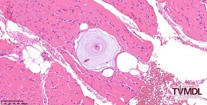
These parasites are often widely distributed throughout all skeletal muscle. Since they can be scattered through the tissue, submission of multiple biopsy specimens is recommended to increase the chance of collecting a diagnostic sample. Histologically, organisms can be found in skeletal and cardiac muscle and may be accompanied by pyogranulomatous inflammation. The parasites are often surrounded by multiple layers of mucopolysaccharide, creating a characteristic “onion-skin” cyst,
Read more by clicking on the link below:
Canine hepatozoonosis diagnosed via skeletal muscle biopsy
Share This Article
Free Membership
Enjoyed this article?
There's a lot more where that came from.
Join 50,000+ veterinary professionals who get free RACE-approved CE, weekly clinical updates, and the most talked-about veterinary magazine in the profession — all completely free.
Join Vet Candy Free →No credit card. No catch. Just everything veterinary.