

The case of the painful Boston terrier
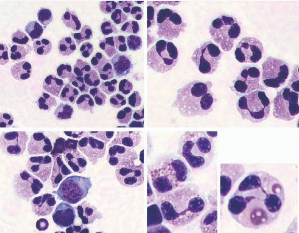
Oreo is a 7-month-old male Boston Terrier presented to a neurologist with a history of episodic back pain. A magnetic resonance imaging (MRI) was performed with results being unremarkable.A sample of cerebral spin fluid (CSF) was collected from the lumbar cistern, revealing an inflammatory profile containing 1,575 nucleated cells and 50 red blood cells.
A good rule of thumb for interpretation is to find a cell that represents the normal expected appearance. Unfortunately, there was not a perfect neutrophil to be found for comparison in this sample, but overall these appear consistent with eosinophils. All of the granulocytes present are morphologically similar, there is more distinct pink granules in many of them, and the bilobed “Mickey Mouse ears” nuclear morphology is more commonly present in eosinophils, specifically.
Possible differential diagnoses for eosinophilic pleocytosis in dogs include broad categories such as non-specific acute inflammatory (SRMA, idiopathic), parasitic, allergic, & neoplastic. Specific infectious causes reported include protozoa (toxoplasmosis, neosporosis), Protothecosis, Cryptococcosis, viral (distemper, rabies), cuterebriasis, as well as aberrant migration of internal parasites.
Read more: