What you need to know about glaucoma in pets
What is glaucoma and how does it develop?
Glaucoma is a group of diseases that results in increased intraocular pressure (IOP). Increased IOP is painful and causes damage to the retina and optic nerve, which can lead to irreversible blindness. It is a leading cause of blindness in animals and people.
Glaucoma develops due to a disturbance in the flow of fluid within and out of the eye. Aqueous humor is the fluid inside the front chamber of the eye, which is produced by another part of the eye called the ciliary body. Aqueous humor provides nutrition to the lens and cornea. In dogs and cats, it is predominantly drained out of the eye by the iridiocorneal angle (ICA). When this angle, or drain, is obstructed for any reason, aqueous humor builds up inside the eye and results in increased IOP. This is similar to your kitchen sink getting clogged with food, and the water backing up in the sink.
There are 2 classifications of glaucoma, primary and secondary. Primary glaucoma is inherited and breed-related, so affected animals have an abnormal formation of their ICA, or drain, at birth. Predisposed breeds include the Cocker Spaniel, Basset Hound, Beagle, Great Dane, Siberian Husky, Jack Russell Terrier, Samoyed, Chow Chow, and Chinese Shar Pei. Primary glaucoma is a bilateral condition, but both eyes are not typically affected at the same time. Once one eye is affected, the other eye is usually affected within 2 years; however, with preventative medical therapy, this time interval can be extended. Secondary glaucoma is acquired, and affected animals have a secondary disease or injury causing increased IOP. It is typically unilateral, and causes of secondary glaucoma include but are not limited to anterior lens luxation (movement of the entire lens into the front of the eye), uveitis (inflammation inside the eye), intraocular masses, hyphema (blood inside the eye), and cataracts.
How is glaucoma diagnosed?
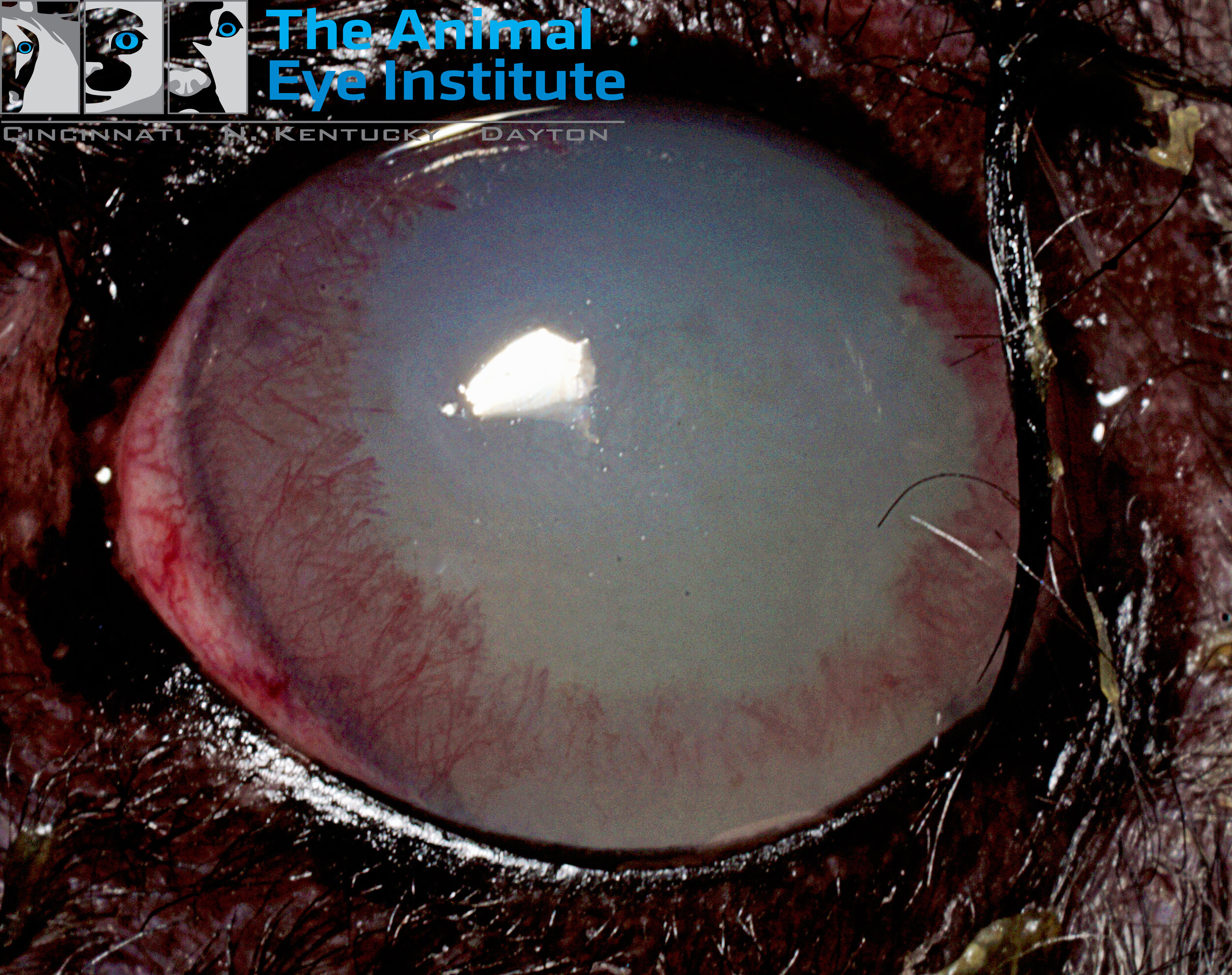
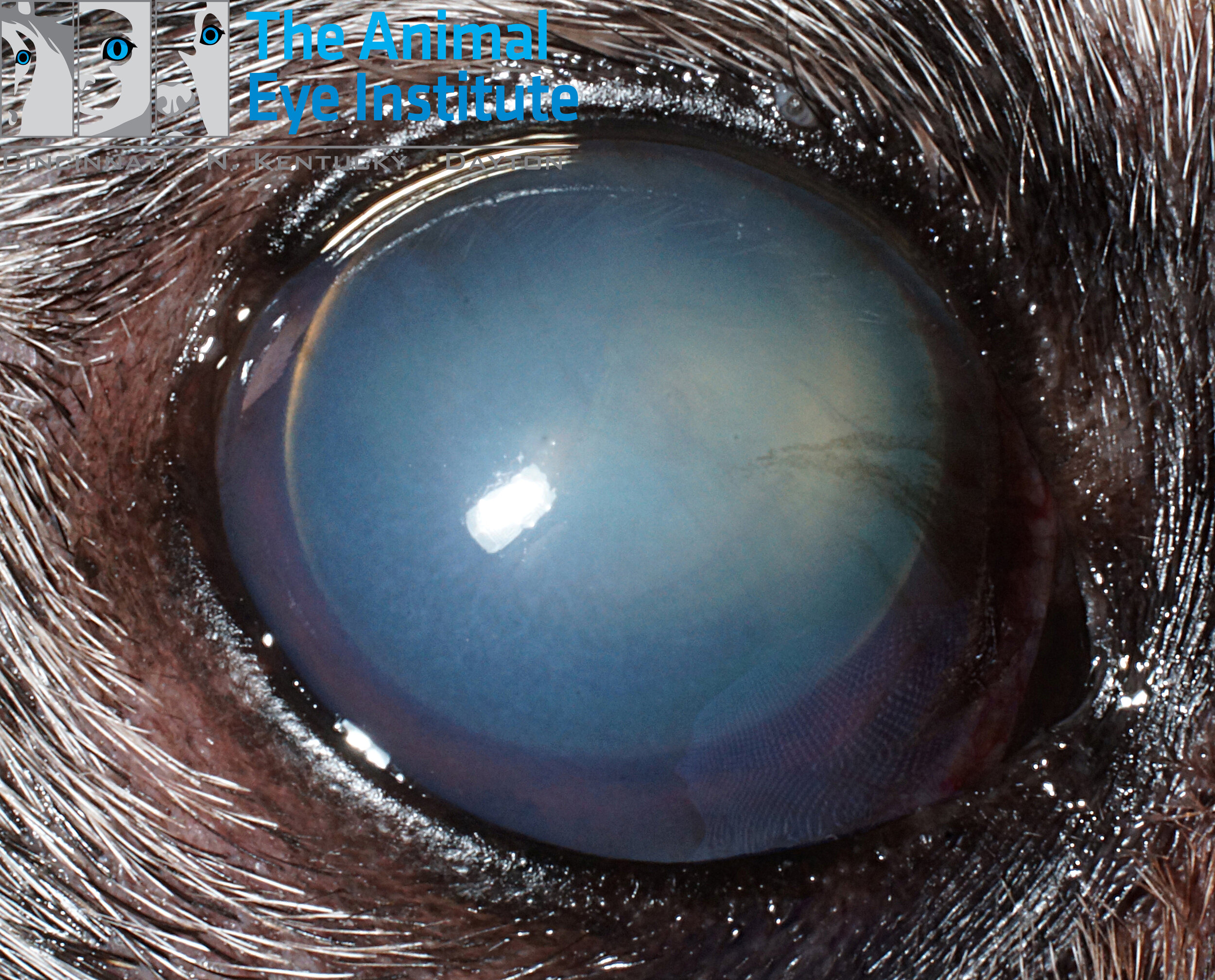
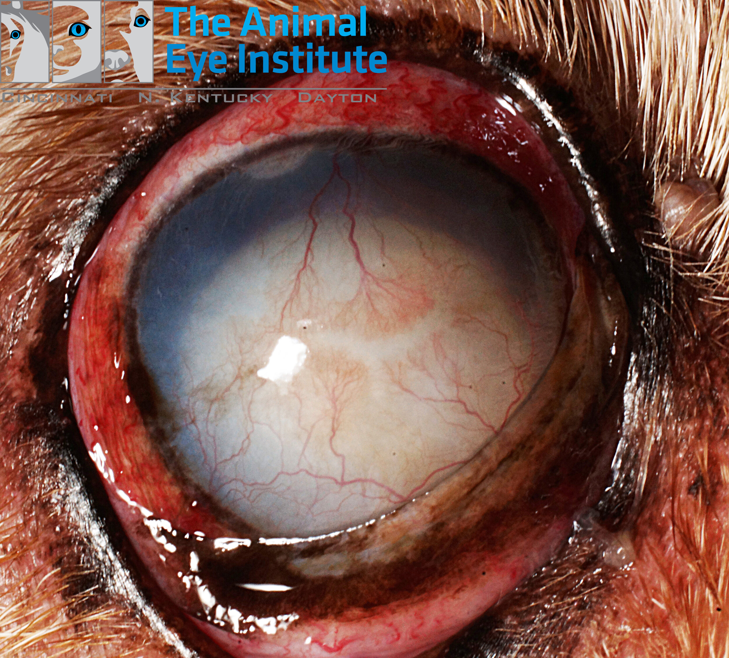
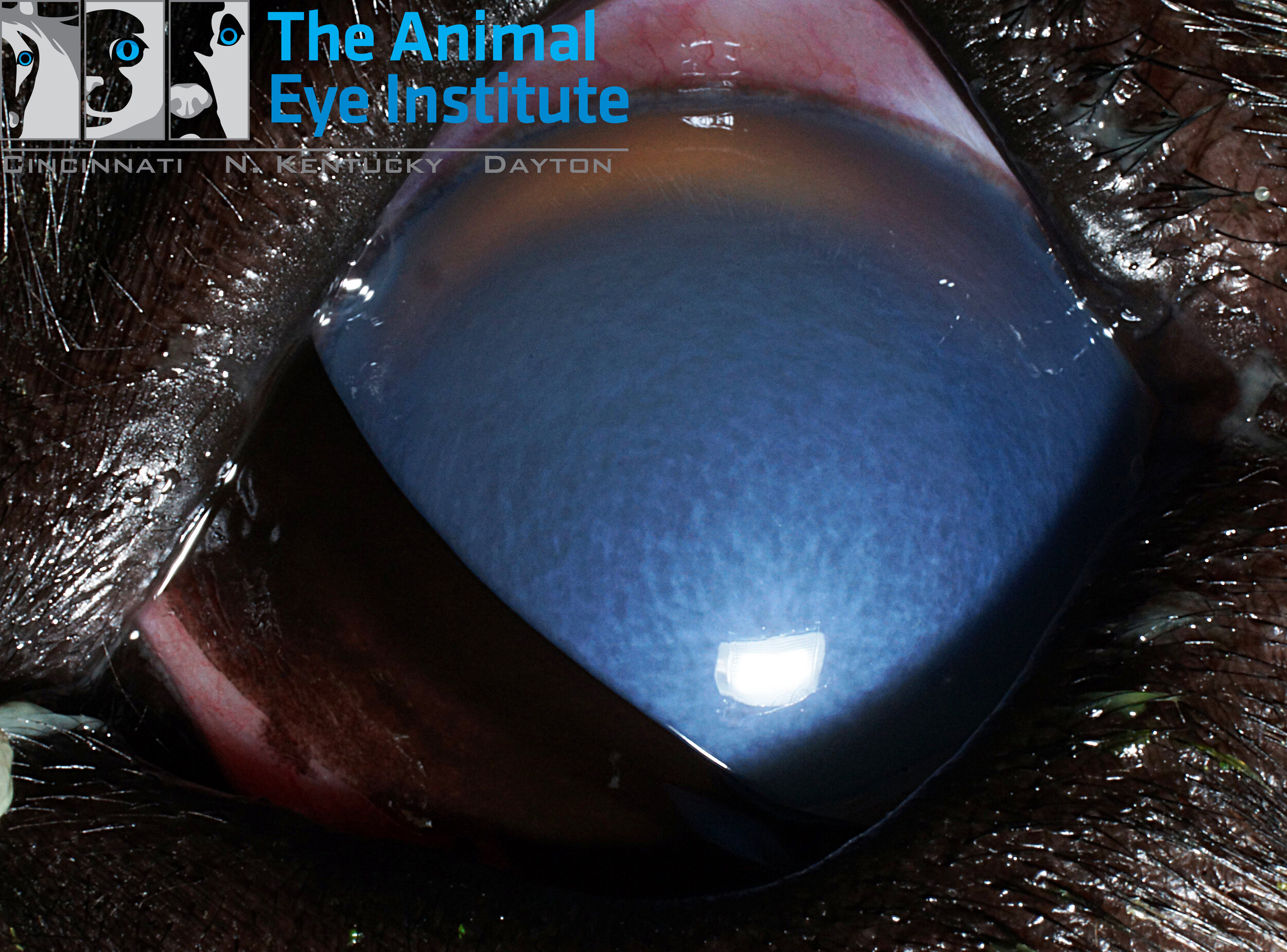
Glaucoma is diagnosed by using a specialized instrument called a tonometer to measure IOP. A diagnosis is supported by a complete ophthalmic examination. Clinical signs of glaucoma include redness, cloudiness, tearing, loss of vision, an enlarged eye, lethargy, and inappetance. It is a painful disease and can be compared to a migraine headache in humans.
In some cases, the drainage structure of the eye can be examined using a special lens (gonioscopy), which can help classify the type of glaucoma. Understanding whether a dog has primary or secondary glaucoma is helpful, as this can help direct whether treating the unaffected eye would be beneficial.
How is glaucoma treated?
Acute glaucoma is considered an emergency! There is no cure for glaucoma, and it can be difficult to control. There are both medical and surgical treatment options. There are several different topical medications with different mechanisms of action that can be used to control glaucoma. If these medications fail, surgical options are available. Surgical options include the use of a laser to damage cells of the ciliary body which produces aqueous humor and surgical placement of implants that drain excess aqueous humor from the eye.
Due to the aggressive nature of the disease, some animals lose vision despite treatment. There are still options for blind and painful eyes, including eye removal and placement of an intraocular prosthesis.
What is the prognosis of glaucoma?
Prognosis is dependent on early detection and response to treatment. Immediate evaluation by a board certified veterinary ophthalmologist is key in providing the best possible outcome. It is a disease that requires lifelong treatment and monitoring by a veterinary ophthalmologist.